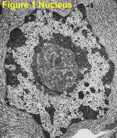
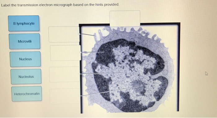
41 label the transmission electron micrograph of the nucleus
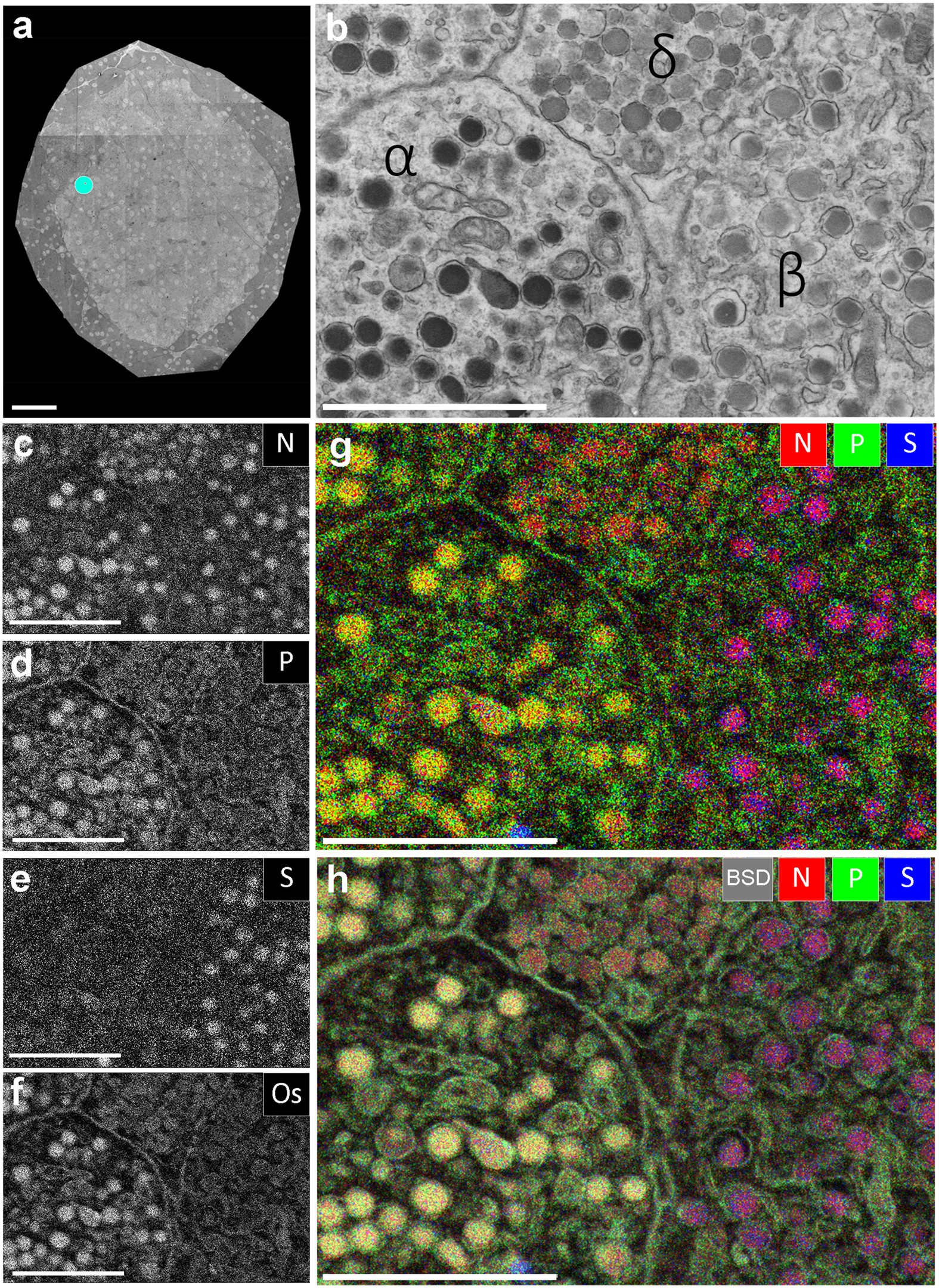
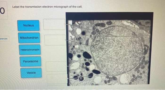
Transmission electron microscopy of the cell nucleus Transmission electron microscopy of the nucleus: This image is an electron spectroscopic imaging (also known as energy filtered transmission electron microscopy) image of a mouse 10T1/2 fibroblast nucleus. Quantitative distributions of phosphorus, which highlights DNA and RNA due to its inherent high phosphorus content were false-coloured green. Solved Label the transmission electron micrograph of the - Chegg Label the transmission electron micrograph of the cell. 0 Nucleus rences Mitochondrion Heterochromatin Peroxisome Vesicle ULAR bumit Click and drag each label into the correct category to indicate whether it pertains to the cytoplasm or the plasma membrane.
Quantum theory of electronic excitation and sputtering by transmission ... Abstract. Many computational models have been developed to predict the rates of atomic displacements in two-dimensional (2D) materials under electron beam irradiation. However, th

Label the transmission electron micrograph of the nucleus
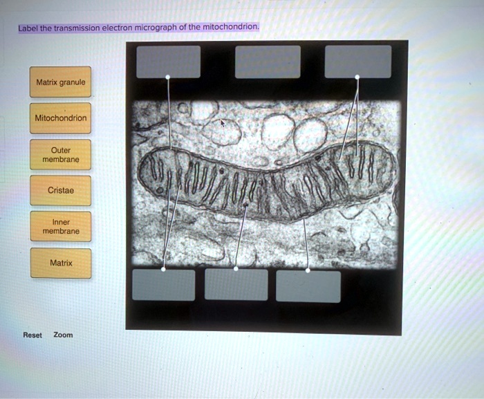
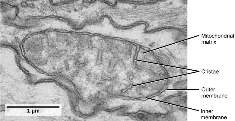
Looking at the Structure of Cells in the Microscope Determining the detailed structure of the membranes and organelles in cells requires the higher resolution attainable in a transmission electron microscope. Specific macromolecules can be localized with colloidal gold linked to antibodies. Three-dimensional views of the surfaces of cells and tissues are obtained by scanning electron microscopy. Eukaryotic Cells | Biology I - Lumen Learning The centrosome is a region near the nucleus of animal cells that functions as a microtubule-organizing center. It contains a pair of centrioles, two structures that lie perpendicular to each other. ... This transmission electron micrograph shows a mitochondrion as viewed with an electron microscope. Notice the inner and outer membranes, the ... APR 2 Flashcards | Quizlet Label the transmission electron micrograph of the nucleus. Place the following cytoplasmic structures in the appropriate structural category. Non-membrane-bound organelle 1. Ribosome 2. Centrosome Membrane-bound organelle 1. Golgi apparatus 2. Mitochondrion 3. Lysosome 4. Peroxisome 5. Rough endoplasmic reticulum Cytoskeleton 1.
Label the transmission electron micrograph of the nucleus. The Transmission Electron Microscope | CCBER - UC Santa Barbara Transmission electron microscopes (TEM) are microscopes that use a particle beam of electrons to visualize specimens and generate a highly-magnified image. TEMs can magnify objects up to 2 million times. In order to get a better idea of just how small that is, think of how small a cell is. Virtual EM Micrograph List | histology - University of Michigan 021. Plasma Cell: This electron micrograph shows a typical secretory cell, a plasma cell, which secretes immunoglobulin protein. Many of the major types of cellular organelles are visible in this image. In the nucleus, areas of euchromatin and heterochromatin can easily be identified. Virtual Slide. Labeling the Cell Flashcards | Quizlet outside the cell wall Label the transmission electron micrograph of the cell. Label the transmission electron micrograph of the mitochondrion. Label the transmission electron micrograph of the nucleus. Students also viewed Chapter 3 Worksheet 50 terms Images sonjamilosavljevic Recent flashcard sets Unit 4 Forces of nature 16 terms 4.3 Eukaryotic Cells - Biology 2e | OpenStax Unlike prokaryotic cells, eukaryotic cells have: 1) a membrane-bound nucleus; 2) numerous membrane-bound organelles such as the endoplasmic reticulum, Golgi apparatus, chloroplasts, mitochondria, and others; and 3) several, rod-shaped chromosomes. Because a membrane surrounds eukaryotic cell's nucleus, it has a "true nucleus.".
Electron microscope - Wikipedia An electron microscope is a microscope that uses a beam of electrons as a source of illumination. They use electron optics that are analogous to the glass lenses of an optical light microscope. As the wavelength of an electron can be up to 100,000 times shorter than that of visible light, electron microscopes have a higher resolution of about 0.1 nm, which compares to about 200 nm for light ... Animal cells - Cell structure - AQA - GCSE Biology (Single Science ... Animal cells have a basic structure. Below the basic structure is shown in the same animal cell, on the left viewed with the light microscope, and on the right with the transmission electron ... Electron microscopes - Cell structure - Edexcel - BBC Bitesize the transmission electron microscope (TEM) is used to examine thin slices or sections of cells or tissues the scanning electron microscope (SEM) has a large depth of field so can be used to ... 10.1: Plant Cell Structure and Components - Biology LibreTexts The nucleolus (A) is a condensed region within the nucleus (B) where ribosomes are synthesized. The nucleus is surrounded by the nuclear envelope (C). Just oustide the nucleus, the rough endoplasmic reticulum (D) is composed of many layers of folded membrane. Image from the public domain, via Wikimedia Commons, labels added by Maria Morrow.
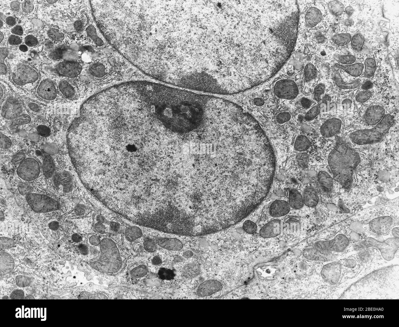
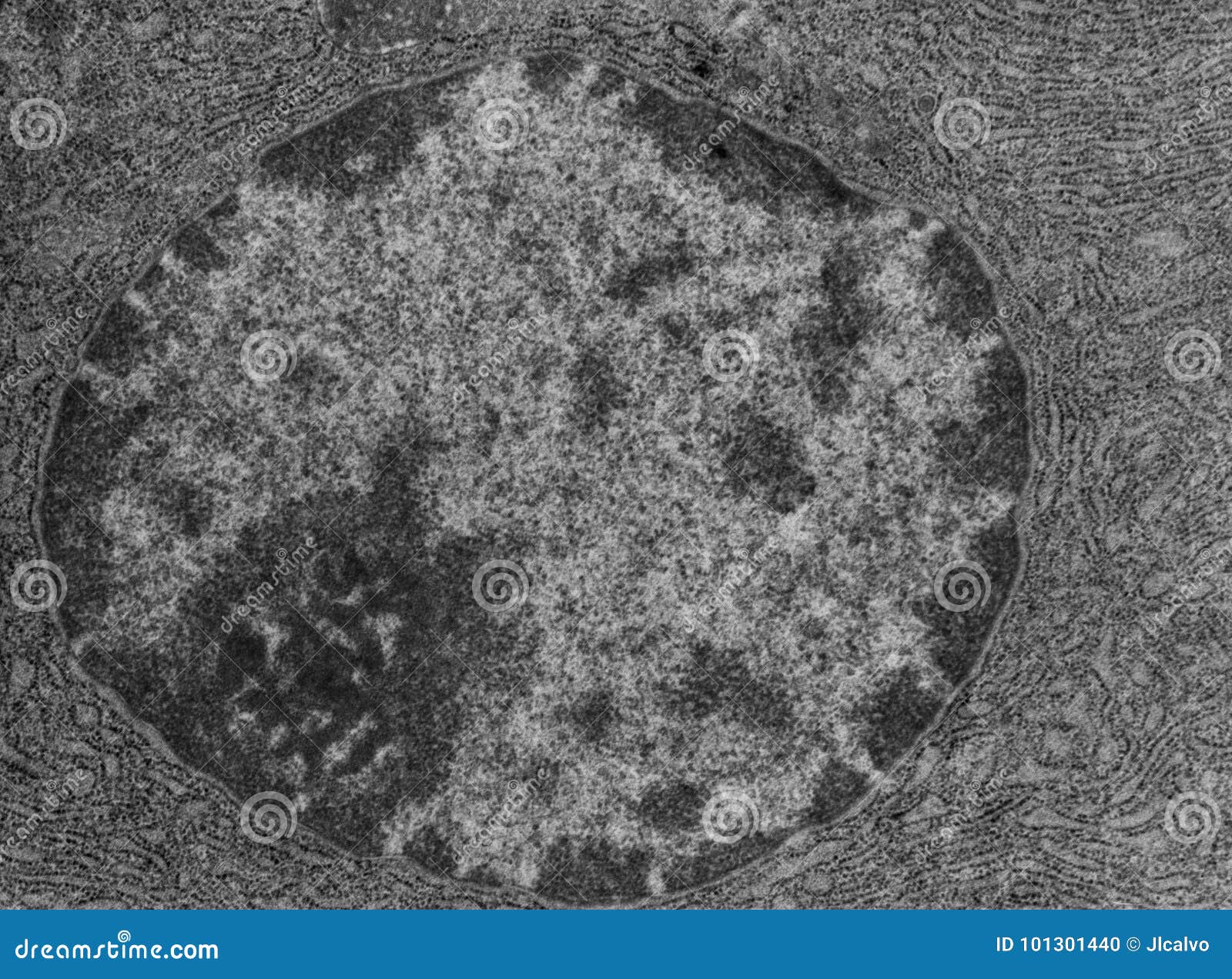
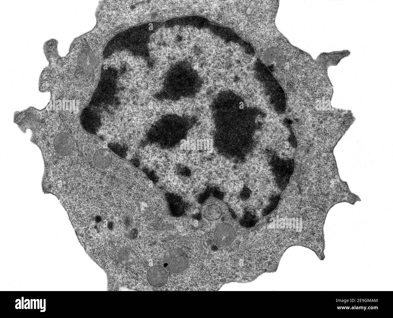
Cell nucleus and English letters - PMC - National Center for ... Neutrophils have nuclei, which are often segmented nucleus; under normal circumstances, they have 2-4 sub leaf nucleus. While the slightly naive cells can be band form nucleus, the transitional nuclei are hyper segmentation and the configuration of the nucleus changes in a random, natural, and disordered manner, forming some special ... The Cell: The Histology Guide - University of Leeds This picture shows an electron micrograph of a nucleus. The short white arrows are pointing to nuclear pores. Note the appearance of eu- and heterochromatin, and the nucleolus. Heterochromatin stains more densely than euchromatin, but they are both forms of chromatin. Chromatin is the name for the diffuse granular mass of DNA found in ... 2.3: Eukaryotic Cell: Structure and Function - Biology LibreTexts The nucleus. Typically, the nucleus is the most prominent organelle in a cell (see figure below) when viewed through a microscope. The nucleus (plural = nuclei) houses the cell's DNA. Let's look at it in more detail. Figure 4. The nucleus stores chromatin (DNA plus proteins) in a gel-like substance called the nucleoplasm. Solved Label the transmission electron micrograph of the - Chegg Question: Label the transmission electron micrograph of the nucleus. Nuclear envelope Nucleolus Nucleus Heterochromatin Reset Zoom Show transcribed image text Expert Answer 100% (25 ratings) Transcribed image text: Label the transmission electron micrograph of the nucleus. Nuclear envelope Nucleolus Nucleus Heterochromatin Reset Zoom
View Image
Electron Micrographs - University of Oklahoma Health Sciences Center Below is a collection of electron micrographs with labelled subcellular structures that you should be able to identify. Also, be sure to observe any electron micrographs which are made available in the laboratory by the instructor. You should concentrate on the similarities in form that permit

Solved Mitochondrion Nucleus Vesicle Peroxisome | Chegg.com
Solved Label the transmission electron micrograph based on - Chegg Expert Answer. 100% (1 rating) So …. View the full answer. Transcribed image text: Label the transmission electron micrograph based on the hints provided Mitochondrion Heterochromatin Plasma cell Nucleus Rough endoplasmic reticulum Nucleolus.
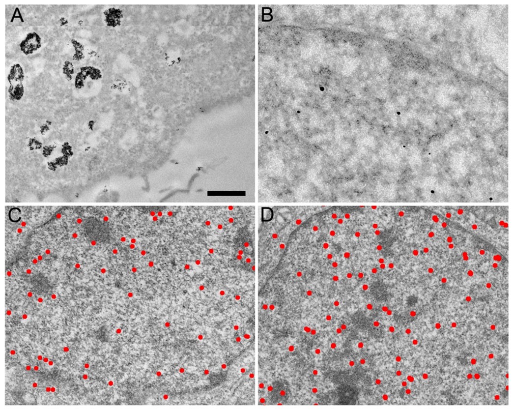
Live cell immunogold labelling of RNA polymerase II ...
Atomic-scale in situ observation of electron beam and heat induced ... Figure 1 shows the high-angle annular dark-field scanning transmission electron microscopy (HAADF-STEM) images of the as-synthesized amorphous Ge solids and hollow nanoparticles. The average diameters of the solid and hollow Ge nanoparticles are 7.3 ± 0.4 nm and 9.3 ± 0.4 nm, respectively. ... (Z = 32) nucleus and its amorphous structure. It ...

False Colour Transmission Electron Microscope (TEM ...
3.3 Eukaryotic Cells - Concepts of Biology - 1st Canadian Edition The centrosome is a region near the nucleus of animal cells that functions as a microtubule-organizing center. It contains a pair of centrioles, two structures that lie perpendicular to each other. ... Figure 3.17 This transmission electron micrograph shows a mitochondrion as viewed with an electron microscope. Notice the inner and outer ...

587 Transmission Electron Micrograph Images, Stock Photos ...
Scanning electron microscopy of nuclear structure - PubMed Abstract. Accessing internal structure and retaining relative three dimensional (3D) organization within the nucleus has always proved difficult in the electron microscope. This is due to the overall size and largely fibrous nature of the contents, making large scale 3D reconstructions difficult from thin sections using transmission electron ...
Transmission Electron Micrograph (TEM) of a cell, showing the ...
Detail of the perinuclear GA of myotubes. (a) Transmission electron ... Previous electron microscopy (EM) studies of myotubes showed that five to seven Golgi cisternae align as a perinuclear belt along the NE, with the cis-Golgi facing towards the NE at a constant ...

Transmission electron-micrograph of the tubal tonsil showing ...
APR 2 Flashcards | Quizlet Label the transmission electron micrograph of the nucleus. Place the following cytoplasmic structures in the appropriate structural category. Non-membrane-bound organelle 1. Ribosome 2. Centrosome Membrane-bound organelle 1. Golgi apparatus 2. Mitochondrion 3. Lysosome 4. Peroxisome 5. Rough endoplasmic reticulum Cytoskeleton 1.

3.3 Eukaryotic Cells – Concepts of Biology – 1st Canadian Edition
Eukaryotic Cells | Biology I - Lumen Learning The centrosome is a region near the nucleus of animal cells that functions as a microtubule-organizing center. It contains a pair of centrioles, two structures that lie perpendicular to each other. ... This transmission electron micrograph shows a mitochondrion as viewed with an electron microscope. Notice the inner and outer membranes, the ...

Specific, Sensitive, High-Resolution Detection of Protein ...
Looking at the Structure of Cells in the Microscope Determining the detailed structure of the membranes and organelles in cells requires the higher resolution attainable in a transmission electron microscope. Specific macromolecules can be localized with colloidal gold linked to antibodies. Three-dimensional views of the surfaces of cells and tissues are obtained by scanning electron microscopy.

Drug-Carrying Capacity and Anticancer Effect of the Folic ...

2,034 Nucleolus Images, Stock Photos & Vectors | Shutterstock

Label-Free Dynamic Imaging of Chromatin in Live Cell Nuclei ...
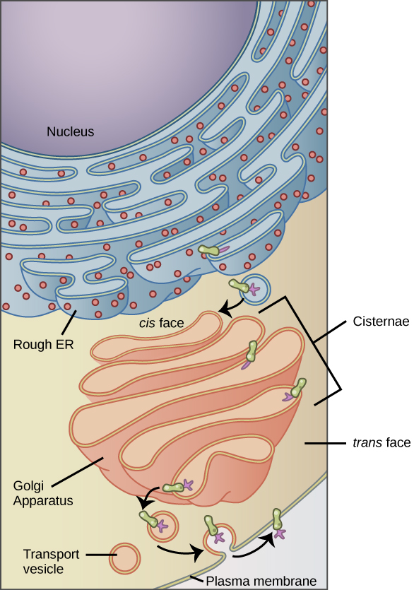
4.4: The Endomembrane System and Proteins - Biology LibreTexts
Electron Micrographs

The Phosphatidylcholine Transfer Protein Stard7 is Required ...

A&P Unit 2 Exam Flashcards | Quizlet

transmission electron micrograph of light cells showing ...
Solved Label the transmission electron micrograph based on ...

Cell Structure & Mitosis Visual Lab - ppt download
SOLVED: I dont know about this one Label the transmission ...
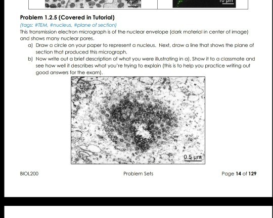
Solved Problem 1.2.5 (Covered in Tutorial) (tags: #TEM ...

Biology, The Cell, Cell Structure, The Endomembrane System ...

IMAGE GALLERY — Columbia Nano Initiative
Nucleus and nucleolus, TEM stock photo. Image of cytology ...
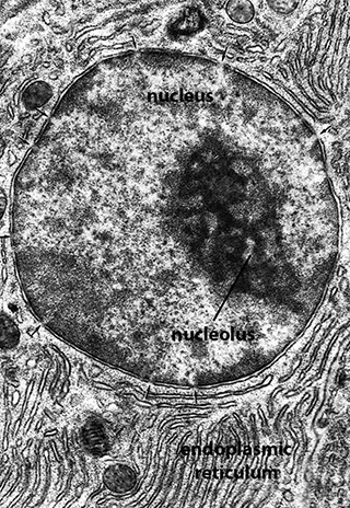
BIOL 230 Lecture Guide - Electron Micrograph of a Nucleus

Transmission electron microscope Black and White Stock Photos ...

Structure and Functions of the Dentin-Pulp Complex | Pocket ...

Labeling the Cell Flashcards | Quizlet

Influenza virus genome reaches the plasma membrane via a ...
Multi-color electron microscopy by element-guided ...
What is a diagram of a plant and animal cell under an ...

Eukaryotic Cells Under the Microscope (2.1.6) | OCR AS ...

File:Gemmata endomembrana2.png - Wikimedia Commons

Cell Micrographs | BioNinja
3.3 Eukaryotic Cells – Concepts of Biology – 1st Canadian Edition

TEM of animal cell - Stock Image - G450/0055 - Science Photo ...

2.3.3 Identify structures from electron micrographs of liver ...

Nanomaterials | Free Full-Text | A Guide for Using ...
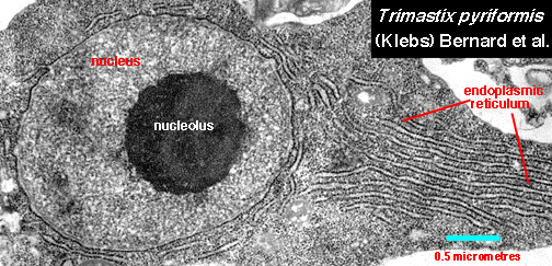
PID - Trimastix Appearance
Solved Label the transmission electron micrograph of the ...
Transmission electron microscope (TEM) micrograph showing a ...
Post a Comment for "41 label the transmission electron micrograph of the nucleus"